Fig. 1.
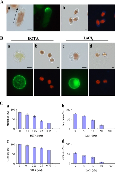
Calcium influx plays an important role in the establishment of cell polarity in monospores. (A) The polarized F-actin and renascent cell wall synthesis in monospores incubated in ESL for 3 h after release from the gametophyte. The polarized F-actin accumulated in the front of the migrating monospores (a) and the renascent cell wall synthesis in migrating monospores (b). The arrow in (a) indicates the direction of migration of the monospore. Left and right photographs in each panel show bright-field and fluorescent images, respectively. Scale bars=5 μm. (B) Effects of calcium chelator (EGTA) and calcium channel blocker (LaCl3) on the F-actin organization and renascent cell wall synthesis. Freshly released monospores incubated with 1 mM EGTA (a, b) and 100 μM LaCl3 (c, d) for 3 h, which indicate the symmetrical organization of F-actin (a, c) and no renascent cell wall synthesis (b, d). Upper and lower photographs in each panel show bright-field and fluorescent images, respectively. Scale bars=5 μm. (C) Dose-dependent inhibition of the motility and development of monospores by calcium chelator and channel blocker. Freshly released monospores incubated with an increasing concentration of EGTA (a, c) and LaCl3 (b, d) for 3 h (a, b) and 24 h (c, d). Columns and vertical bars represent the mean and SD, respectively (n=3).

