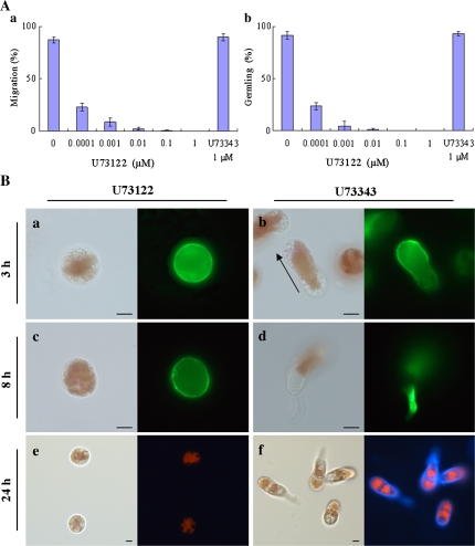
Fig. 2.
Involvement of PLC activity in the establishment of cell polarity in monospores. (A) Effects of PLC inhibitor on the motility and development of monospores. Freshly released monospores incubated with an increasing concentration of U73122 from 0.1 nM to 1 μM and 1 μM U73343 for 3 h (a) and 24 h (b). Columns and vertical bars represent the mean and SD, respectively (n=3). (B) Effects of PLC inhibitor on the F-actin organization and renascent cell wall synthesis. Freshly released monospores incubated with 1 μM U73122 (a, c, e) and 1 μM U73343 (b, d, f) for 3 h (a, b), 8 h (c, d), and 24 h (e, f). The organization of F-actin in U73122 and U73343 treated monospores are indicated in (a, c) and (b, d). Renascent cell wall synthesis in U73122 and U73343 treated monospores are indicated in (e) and (f). Arrow in (b) indicates the direction of migration of the monospore. Left and right photographs in each panel show bright-field and fluorescent images, respectively. Scale bars=5 μm.

