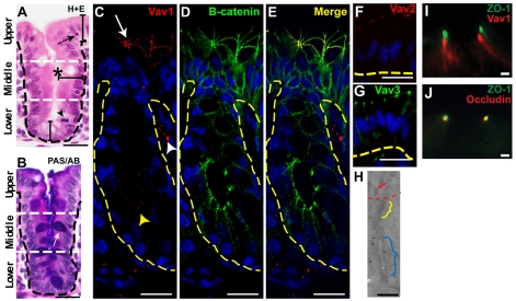
Fig. 1.
Vav proteins are expressed in upper zone enterocytes of the mouse cecum and localize near adherens junctions. (A,B) Sections of WT adult mouse cecum stained with hematoxylin and eosin (H+E) (A) and PAS/alcian blue (B). A single crypt-surface epithelial unit is shown. Black dashed lines indicate the basal epithelial surface. White dashed lines demarcate three epithelial zones. In A, the asterisk denotes the crypt lumen, the arrowhead indicates an M-phase cell in the lower zone and the arrow denotes an apoptotic body in the upper zone. In B, the white arrow denotes a goblet cell (purple). (C-E) Sections of a WT mouse cecum double-labeled with rabbit anti-Vav1, Alexa Fluor 594-conjugated donkey anti-rabbit (red), mouse anti-β-catenin, Alexa Fluor 488-conjugated sheep anti-mouse (green) antibodies and bis-benzimide. Vav1 protein expression was most robust in upper zone enterocytes (arrow; the tangential orientation shows a crypt orifice). The white and yellow arrowheads indicate cells with Vav protein expression in the mesenchyme and crypt epithelium, respectively. (F,G) Sections of WT upper zone cecal enterocytes labeled with anti-Vav2 (F, red) and anti-Vav3 (G, green) antibodies. The yellow dashed lines in C-G indicate the epithelial-mesenchymal junction. (H) Immunogold labeling for Vav1 in an apical junctional complex from a WT upper zone enterocyte. The red arrow denotes microvilli. The yellow and blue brackets indicate tight and adherens junctions, respectively. Vav1 was detected near adherens junctions. (I,J) Sections of a WT upper zone cecal enterocyte double-labeled with anti-ZO-1 (I, green) and anti-Vav1 (I, red) or anti-ZO-1 (J, green) and anti-occludin (J, red) antibodies. Scale bars: A-G, 15 μm; H-J, 200 nm.

