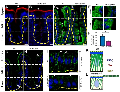
Fig. 2.
Surface enterocytes of Vav1/2/3null mice contain abnormal microtubule networks. (A-D) Sections of cecum from (A,C) WT and (B,D) Vav1/2/3null mice stained with rabbit anti-actin and Cy3-conjugated donkey anti rabbit (red) antibodies and bis-benzimide (blue) (A,B), and FITC-conjugated rabbit anti-α-tubulin (green) and bis-benzimide (C,D). The arrowheads in A and B denote the high level of actin staining in the apex of upper zone enterocytes of both Vav1/2/3null and WT mice. Arrows in C indicate the apical accumulation of polymerized α-tubulin at the cell apex of the lower and middle zones, which is similar to Vav1/2/3null cells in these regions; the arrowhead denotes increased cytoplasmic microtubules in upper zone enterocytes compared with their counterparts in the Vav1/2/3null mice. (E) Insets of boxed regions in C and D from upper and lower zone epithelial cells. (F) Ratio (± s.e.m.) of mean fluorescent intensity of α-tubulin staining in the apical cytoplasm (upper zone/lower zone) of WT and Vav1/2/3null mice. *P<0.01, Student's t-test comparing WT and Vav1/2/3null mice. (G-J) Sections of cecums from WT (G,I) and Vav1/2/3null (H,J) mice stained with rabbit anti-PKCζ and FITC-conjugated donkey anti rabbit (green) antibodies (G,H) and rabbit anti-Dlg1 and FITC-conjugated donkey anti rabbit (green) antibodies (I,J). (K,L) Summary of cellular localization and microtubule networks in upper zone enterocytes in WT and Vav1/2/3null mice. Scale bars: A,C,D,G-J, 15 μm; B, 7.5 μm.

