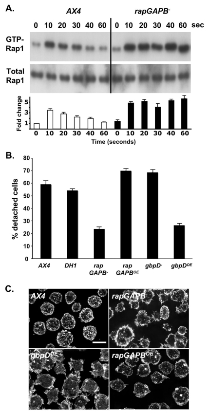
Fig. 2.
RapGAPB is required to regulate Rap1 activity. (A) Western blot showing levels of activated Rap1 (top panel) and total Rap1 (bottom panel). In wild-type cells, maximal increase in levels of active Rap1 was observed after 10 seconds of cAMP stimulation, followed by a steady decrease over 60 seconds of stimulation. In rapGAPB– cells, the maximal increase after 10 seconds of stimulation is slightly higher than that of wild-type cells. In addition, the level of active Rap1 does not decrease over 60 seconds, instead the maximal level is maintained. The level of total Rap1 is equal in all cell lysates. The graph shows fold change in amount of GTP-Rap1 relative to AX4 cells at 0 seconds. (B) Cell-substrate adhesion was measured by counting the number of vegetative cells that detached from a plate after shaking for 20 minutes. Approximately 60% of wild-type cells (AX4 and DH1) detached. Significantly fewer rapGAPB– and gbpDOE cells detached. By contrast, more rapGAPBOE and gbpD– cells detached. (C) Cells were stained with FITC conjugated phalloidin in order to observe F-actin distribution and viewed by deconvolution microscopy. rapGAPB– and gbpDOE cells exhibit a clearly different F-actin distribution and cell morphology compared with wild-type AX4 cells, whereas the rapGAPBOE cells appeared more like the wild type. Scale bar: 10 μm.

