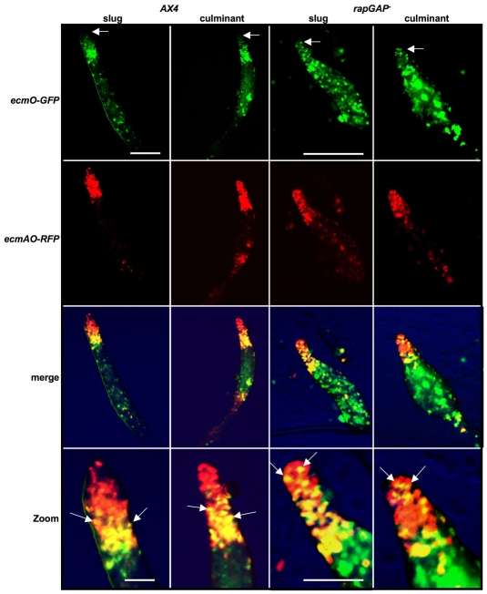
Fig. 5.
Prestalk A and prestalk O cells are intermingled in the rapGAPB– mutant. AX4 and rapGAPB– cells expressing both ecmO-GFP and ecmAO-RFP were developed to the slug and early culminant stages. In developing AX4 cells, ecmO-GFP expression (green) was found mainly in the collar region of slugs and was mostly absent from the tip (arrowed). ecmAO-RFP expression (red) was found throughout the prestalk region of slugs. As a result, the merged image shows orange-yellow cells only in the collar (arrowed in the magnified image) and only red cells in tips of slugs. By contrast, in developing rapGAPB– cells, ecmO-GFP-expressing cells were found in both the collar and tip regions of the slug (arrowed) and ecmAO-RFP expression was found throughout the entire prestalk region. The resulting merged image shows orange-yellow cells in both the collar and tip of the slug (arrowed on the magnified image). A similar defect was observed at the early culminant stage. In AX4 culminants, ecmO-GFP expression was highest in the upper cup and largely absent from the apical tip (arrowed), whereas ecmAO-RFP expression was found in both the collar and apical tip. The merged image shows orange-yellow cells in only the upper cup (arrowed on the magnified image) and only red cells in the apical tip. However, in rapGAPB– culminants, ecmO-GFP and ecmAO-RFP expression were both found in the upper cup and apical tip (arrowed). The merged image shows orange-yellow cells in both the upper cup and apical tip (arrowed in the magnified image). Scale bars: 1 mm and 0.3 mm (zoom panels).

