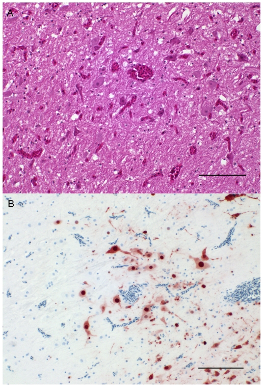
Figure 3. Histopathology and immunohistochemistry of brain from infected ducks.
(A) Brain, Cerebrum; Duck at 5 dpc. Congestion shown by hematoxylin-eosin staining. Bar 100 µm. (B) Brain, Cerebrum; Duck at 6 dpc. Intense intranuclear and intracytoplasmic AIV antigen staining within neurons and neuroglia. Immunohistochemistry. ABC method using anti-NP monoclonal antibody HB65, hematoxylin counterstain. Bar 100 µm.

