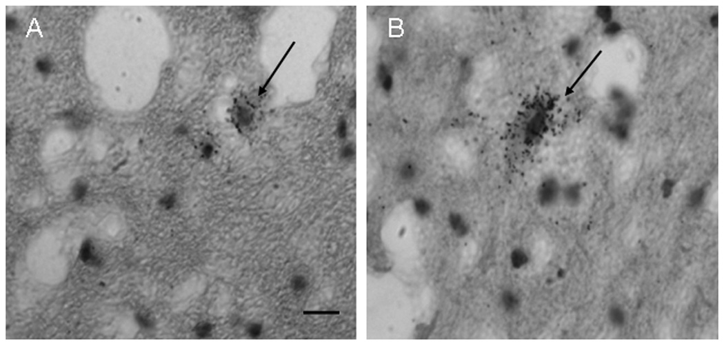
Figure 4.

GAD65 mRNA-labeled neurons (arrows) in the dentate nuclei in a control case of a small cell in A) and a larger cell in B). Note the silver grain GAD65-mRNA-positive labeling throughout both cells. Scale bar in = 20 µm and refers to both A and B.
