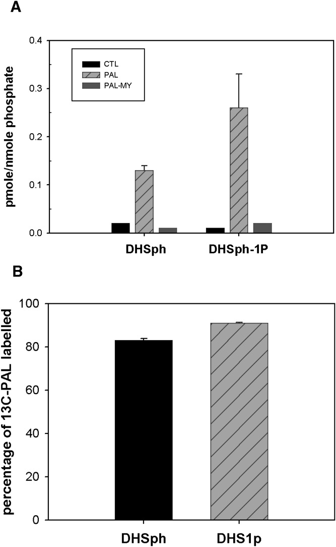
Fig. 1.
PAL increases cellular DHSph and DHSph-1-P. Mouse C2C12 myoblasts were cultured and differentiated into multinucleate myotubes as described in Materials and Methods. A: Cells were treated with 1.25 mM PAL with or without 0.1 μM myriocin. Lipid profiles were determined by LC/MS as described in Materials and Methods. Data are means ± SEM (n = 6) of four experiments. For both DHSph and DHSph1p, CTL versus PAL, P < 0.05; PAL versus PAL-MY, P < 0.05. B: Cells were treated with 1.25 mM uniformly labeled 13C- PAL. The percentage of 13C-PAL labeled in DHSph and DHSph-1-P was determined by LC/MS analysis. Data are means ± SEM (n = 3). CTL, control; MY, myriocin.

