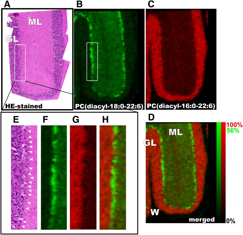
Fig. 6.
Purkinje cells selectively contained a DHA-containing PC. High-magnification IMS at a raster size of 15 μm revealed the Purkinje cell-selective distribution of PC (diacyl-18:0/22:6) in the cerebellum. Both optical observation of HE-stained successive brain sections (A and E) and ion images of DHA-PCs (B and F) clearly suggest that the PC was enriched in the Purkinje cell layer (arrowheads). Interestingly, a complementary distribution of another abundant DHA-PC, PC (diacyl-16:0/22:6), was enriched in the granule layer of the cerebellum (C and G). D: merged image. ML, molecular layer; GL, granule layer; W, white matter. The relative abundance of the two ions is indicated in the color scale bar.

