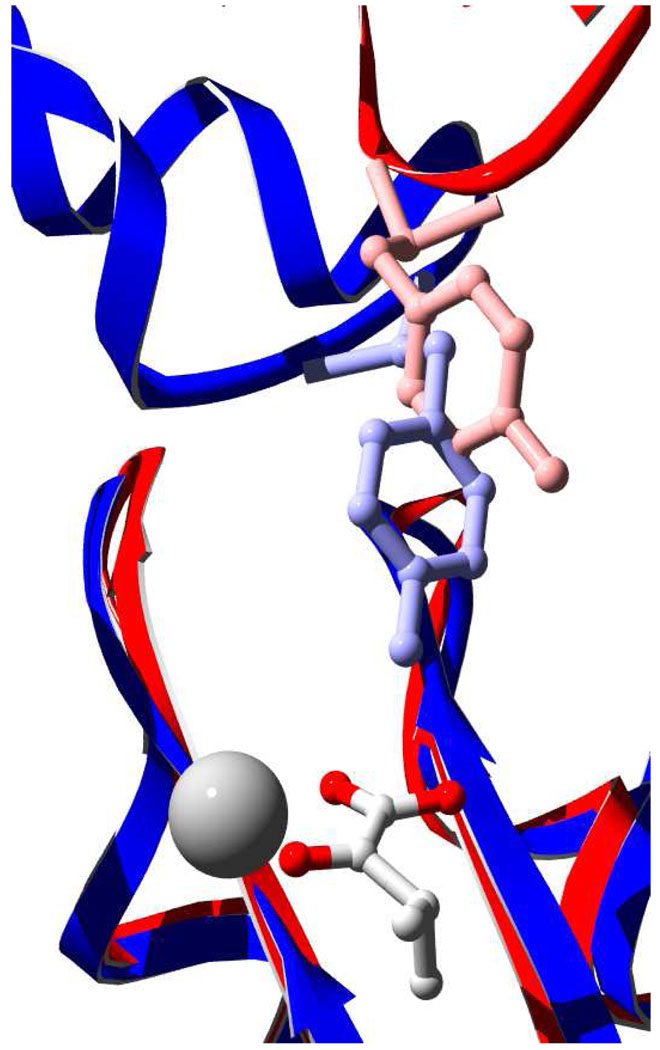
Figure 5.
Superimposition of MtIPMS and DmpG. Swiss-PDB Viewer was used to align the E chain of DmpG (1NMV) (blue) with MtIPMS (1SR9) (red). The alignment included 808 atoms and had an RMS of 1.4Å. α-KIV and a Mg2+ atom are shown to indicate the active site of MtIPMS. MtIPMS Y410 (red) and DmpG Y291 (blue) are depicted as ball and stick representations.

