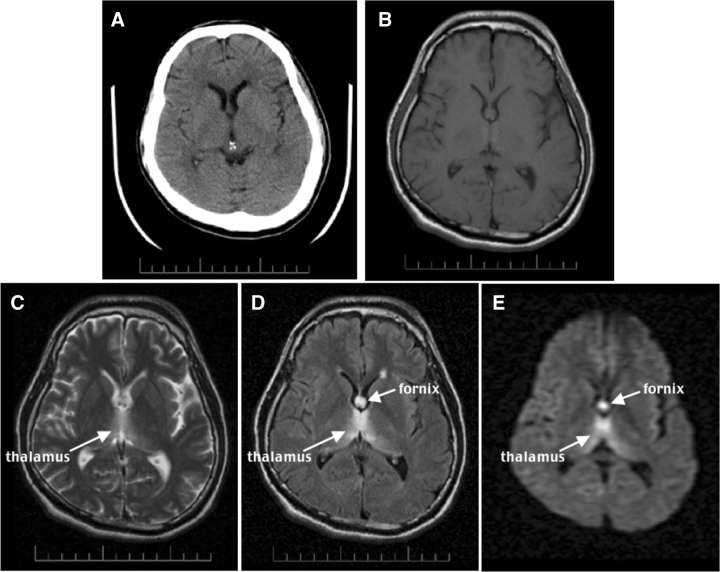
Fig. 1.
CT and MR images of an acute 35-year-old man with schizophrenia and acute nutritional deficiency-induced WE. (A) Axial CT at the level of the lateral ventricles. (B–E) Axial MR images at a similar level to the CT. (B) A proton density-weighted image. (C) A T2-weighted late-echo fast spin echo (FSE) image. (D) A fluid-attenuated inversion recovery (FLAIR) image. (E) A diffusion-weighted image (DWI). Note the hyperintensity of the fornix and thalamus, especially in D and E, less so in C, and lack of lesion conspicuity in A and B.

