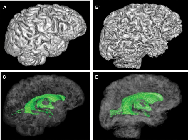
Fig. 3.
Surface rendered brains (top) and rendered ventricular system (bottom, green) of a 59-year-old healthy man (A and C) and a 53-year-old man with WKS (B and D). Note the shrinking of the cortical gyri and widening of the sulci (B) and expansion of the ventricles (D) of the WKS compared with the control (A and C).

