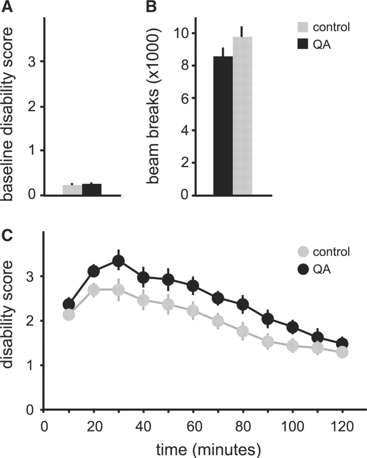
Fig. 8.
Microinjection of kainic acid into the cerebellum following QA lesions in normal mice. Mice with QA lesions (n = 14) are shown in black and saline controls (n = 13) are shown in grey. Results are average values ± SEM. Baseline motor function is shown as motor disability scores (A) or gross motor activity in automated photocell chambers (B). (C) Shows the temporal profile of motor disability due to dystonic motor behaviour after intra-cerebellar microinjection of 50 μg/ml kainic acid in 0.5 μl at time 0. Two-way ANOVA with time as a repeated measure revealed significant effects for time [F(11,275) = 56.9, P < 0.001] and group [F(1,25) = 10.3, P < 0.005]. The interaction between time and group was not significant [F(11,275) = 1.1, P = 0.33].

