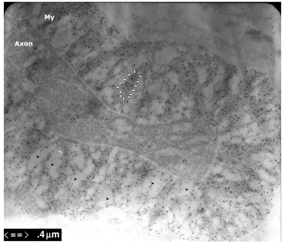
Figure 1.

Immuno-electron microscopy of dermal myelinated nerve fibres. Skin biopsies were fixed for 3 h and embedded in LR white. The ultra-thin sections were stained with antibodies against PMP22 that were conjugated with gold particles. These particles were mainly found in compact myelin, but minimally in axons. Immuno-electron microscopy fixation techniques cannot completely preserve the compact myelin and usually results in vacuolated areas in the compact myelin (arrowheads). To exclude the effect of these vacuoles on our data, we only measured the grain density in the areas with intact myelin (arrow circle).
