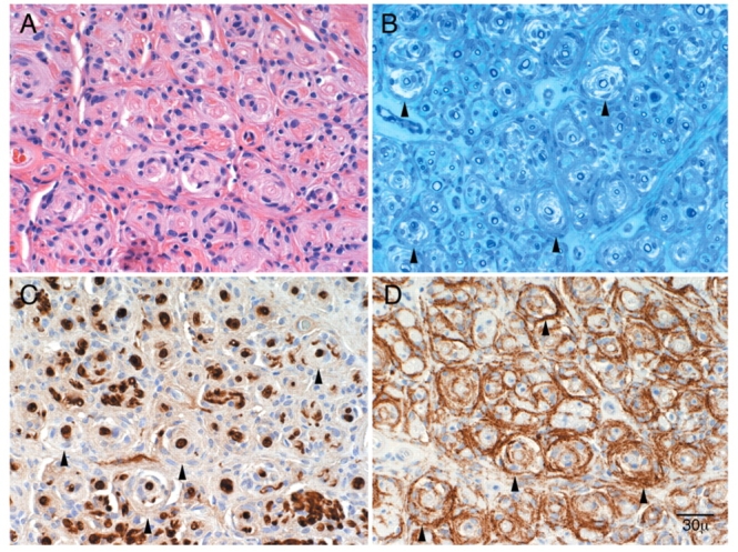
Figure 1.

Pathological features of perineurioma. Patient 1: serial transverse sections: Arrowheads indicate pseudo-onion bulb leaflets. (A) H&E section demonstrates diffuse pseudo-onion bulb formation. (B) Epoxy section at a similar level demonstrates thinly myelinated fibres at the centre of pseudo-onion bulbs. (C) Schwann cell preparation (S-100) demonstrates reactivity of the myelinated fibres at the centre and absence of reactivity of the surrounding pseudo-onion bulbs. (D) Reactivity of pseudo-onion bulb leaflets with epithelial membrane antigen (EMA) confirming these are of perineurial origin. These findings taken together are diagnostic of perineurioma.
