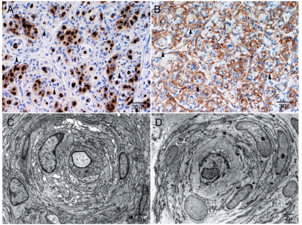
Figure 2.

Immunohistochemistry and ultrastructural features of perineurioma. Patient 3: These pathological features are typical of intraneural perineurioma. In contrast the clinical features of this case are very unusual as this young child has bilateral disease involving multiple nerves over a long distance (see Case 2). (A) S-100 preparation demonstrates reactivity of the myelinated fibres at the centre and absence of reactivity of the surrounding pseudo-onion bulbs (arrowheads). (B) Reactivity of pseudo-onion bulb leaflets with epithelial membrane antigen (EMA) confirming these are of perineurial origin (arrowheads). (C) Electron micrograph of Patient 4 and (D) electron micrograph of Patient 8 demonstrate dense concentrically arranged cellular processes around thinly myelinated axons typical of perineurioma.
