
Fig. 4.
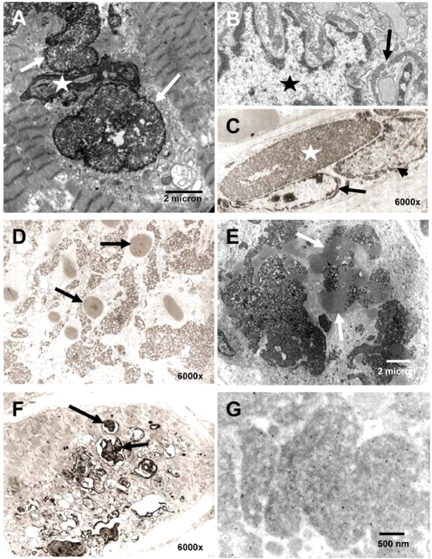
Ultrastructural analysis. (A–C) Localization of aggregates (arrows) in close proximity to a nucleus (star), appearing to indent the nucleus (A). Dense material is seen surrounding the myonucleus (star) as a thin band (B). (D and E) Cytoplasmic bodies (arrows) were found in frequent coexistence with the reducing bodies in all specimens examined. (F) Rimmed vacuoles (arrows) occur in some fibres in RBM (compare Fig. 1D inset). (G) Immuno EM: gold-particles indicate binding of the FHL1 antibody to the major electron-dense aggregates.