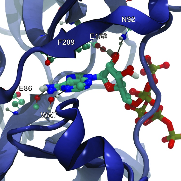
Figure 5.
ATP co-crystallized with TbREL1 (rendered in CPK) superimposed on the AutoGrow-generated ATP docked into the protein receptor (rendered in licorice). Black lines represent hydrogen bonds. The co-crystallized and docked ligands do not overlap in the region of the ATP polyphosphate tail because a magnesium ion, present in the crystal structure but absent in the protein receptor used for docking, coordinates many of the polyphosphate oxygen atoms.

