
Fig. 1. PTPase-defective SHP-2 inhibits growth of EGFRvIII GBM cells.
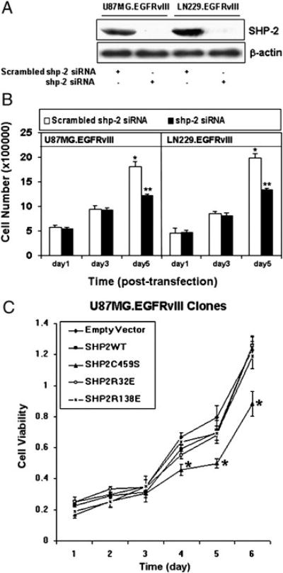
(A) U87MG.EGFRvIII and LN229.EGFRvIII cells were transiently transfected with 20 picomoles of scrambled or shp-2 siRNA for 6 hour and cultured in DMEM plus 10% FBS for 72 h. Equal amounts of protein were loaded for SDS-PAGE gel electrophoresis and western blots were performed with anti-SHP-2 and anti-β-actin antibodies. (B) 20 picomoles of scrambled shp-2 siRNA and shp-2 siRNA were transfected to U87MG.EGFRvIII and LN229.EGFRvIII cells for 6 h and cell number was then counted at the indicated time points. Each column represents the mean ± S.D., n = 4. Data are summarized from four independent experiments giving similar growth profile. A significant difference is presented (* vs **, P<0.05). (C) 4000 cells of distinct EGFRvIII clones expressing different Shp-2 mutants were seeded in 96 well plates and cultured in 10% FBS. The MTT assay was performed at different experimental time points as noted. The data are shown as means ± S.D. of triplicate samples. This is one representative of three separate experiments. Significant differences among the same time points are presented (*, P<0.05).