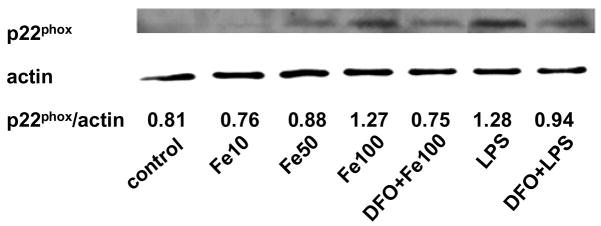
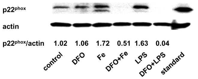
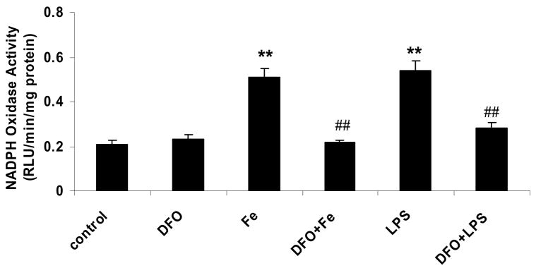
Figure 3. DFO inhibits the LPS or iron-induced increase in p22phox protein and NADPH oxidase activity.
HAEC (panels a and c) or THP-1 cells (panel b) were incubated as described in the legend of Fig. 2, using 5 μg/mL LPS, 100 μmol/L ferric citrate, or 100 μmol/L DFO. For panel a, HAEC were also incubated for 48 hrs with 10 or 50 μmol/L ferric citrate (Fe10 and Fe50, respectively). After incubation, the cells were lysed and assayed for p22phox by Western blot analysis (panels a and b) or NADPH oxidase activity (panel c) as described in Methods. Each Western blot is representative of three independent experiments. Actin was used as loading control, and the numbers below the individual bands in panels a and b indicate the mean ratio of p22phox to actin determined by densitometry (n=3). In panel b, “standard” indicates the THP-1 lysate from Santa Cruz Biotechnology, used as positive control. Panel c: **P<0.01 vs. control; ##P<0.01 vs. Fe or LPS; n=4.


