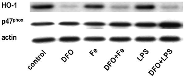
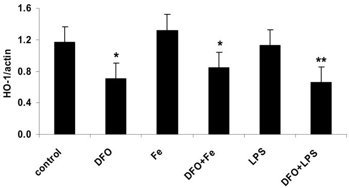
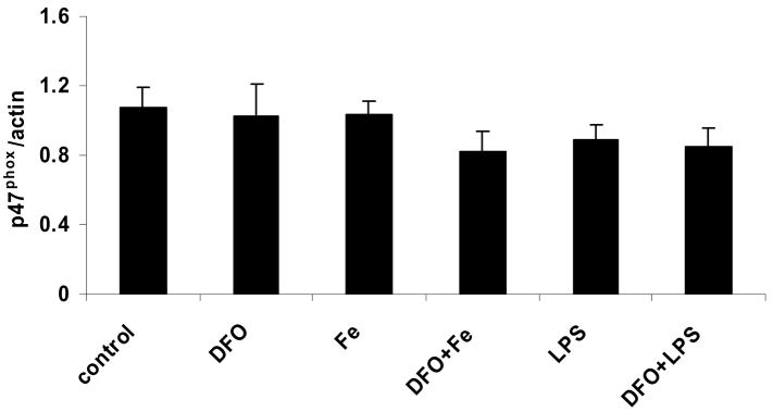
Figure 4. LPS and iron do not increase, but DFO decreases, heme oxygenase-1 protein, whereas p47phox protein is unaffected.
HAEC were incubated as described in the legend of Fig. 2. After incubation, the cells were lysed and assayed for HO-1 and p47phox by Western blot analysis (panel a) as described in Methods. Each Western blot is representative of three (HO-1) or four (p47phox) independent experiments. Actin was used as loading control, and protein levels of HO-1 and p47phox were quantified by densitometry and expressed as the mean ratio of HO-1 to actin (panel b) or p47phox to actin (panel c). Panel b: *P<0.05 and **P<0.01 vs. control; n=3.


