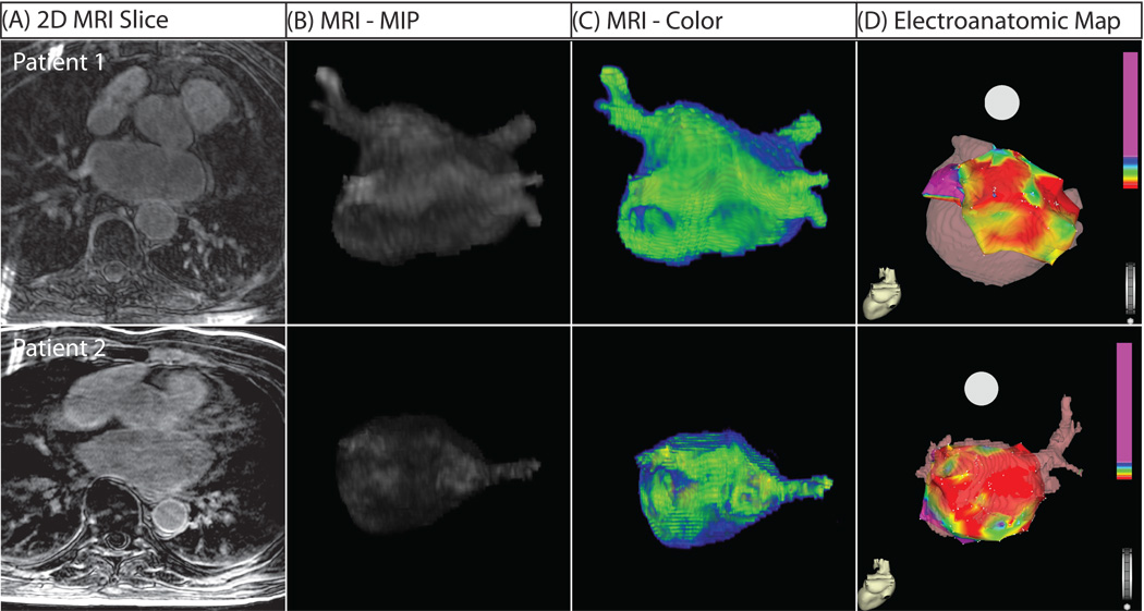
Figure 5. Three-Dimensional MRI Models in Two Patients with Extensive Structural Remodeling.
Both patients shown suffered a recurrence of atrial fibrillation. (A) Two dimensional slice from DE-MRI scan. (B) Segmented DE-MRI reveals large amounts of enhancement in various regions of the LA including anterior wall, posterior wall and septum. (C) MRI images as color 3D models show abnormally enhanced regions (green). (D) EA maps show large regions of electrically non-viable tissue (fibrotic scar) in red interspersed with electrically abnormal tissue (colored).

