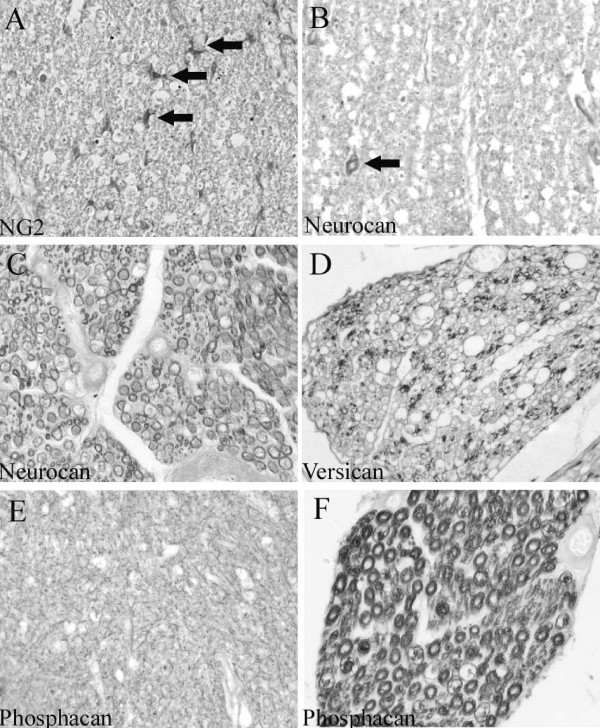
Figure 2.
Normal distribution of NG2, neurocan, versican and phosphacan in the human spinal cord. Transverse sections of control human spinal cords. A: NG2 immunohistochemistry reveals small stellate-shaped cells distributed homogeneously in white matter regions of human spinal cord (arrows). B: In the white matter, neurocan immunoreactivity is observed in the wall of a small blood vessel (arrow). Furthermore, a reticular staining pattern can be seen. C: In a dorsal nerve root, neurocan staining is present in myelin sheaths. D: Versican immunoreactivity is scattered in a dorsal nerve root and can be found in myelin sheaths of small diameter axons. E: Phosphacan immunohistochemistry reveals a fine reticular staining pattern in the gray matter. F: In a dorsal nerve root, phosphacan-immunopositive myelin rings can be observed. (A-F magnification × 320).

