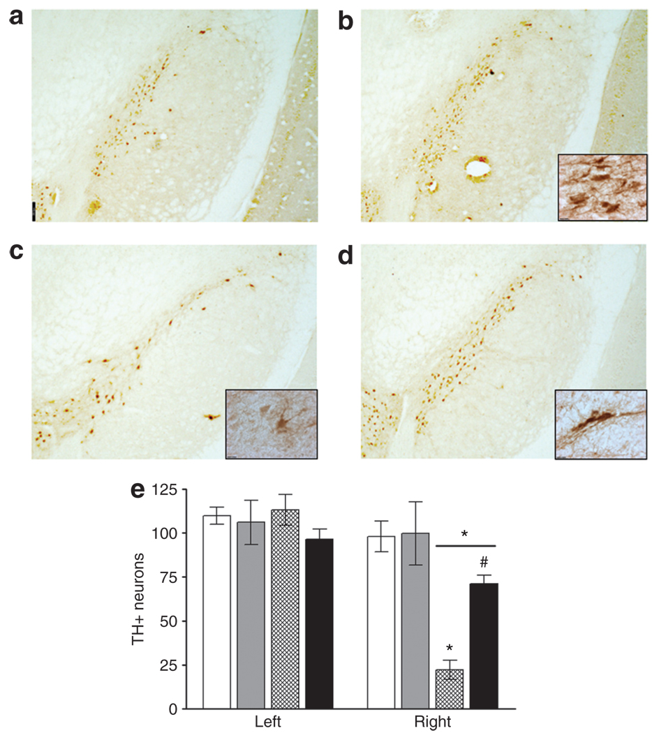
Figure 4. Tyrosine hydroxylase immunoreactive nigral neurons in substantia nigra compacta (SNc) 28 days after infusion of 6- hydroxydopamine (6-OHDA).
White bars, AAV2-GFP/sham; grey bars, AAV2-hIL-10/sham; hatched bars, AAV2-GFP/6-OHDA; and black bars, AAV2-hIL-10/6-OHDA. Striata were infused bilaterally with adeno-associated virus type-2 (AAV2) constructs (1011 vector genomes/20 µl/side) containing a gene for either human interleukin-10 (hIL-10) or green fluorescent protein (GFP). Three days later the right striatum was infused with 20 µg of 6-OHDA or vehicle (sham-lesion). (a) AAV2-GFP/sham; (b) AAV2-h IL-10/sham; (c) AAV2-GFP/6-OHDA; (d) AAV2-h IL-10/6-OHDA. Scale-bar = 300 µm (×2.5 original magnification); (e) mean values are shown for the intact (left) and lesioned (right) SNc at the level of the accessory optic tract (n = 4–6/group). Bars represent total number of tyrosine hydroxylase (TH)-immunoreactive neurons. Right hemisphere data: small *P < 0.001; two-way analysis of variance (ANOVA) followed by Newman–Keuls post-tests, comparisons made with sham-lesioned group. #AAV2-h IL-10/6-OHDA compared with AAV2-GFP/6-OHDA P < 0.01; one-way ANOVA followed by Newman–Keuls post hoc test. Large *P < 0.01 compared to left side within treatment groups; paired two-tailed t-test. Scale bar = 12 µm (×100 magnification).

