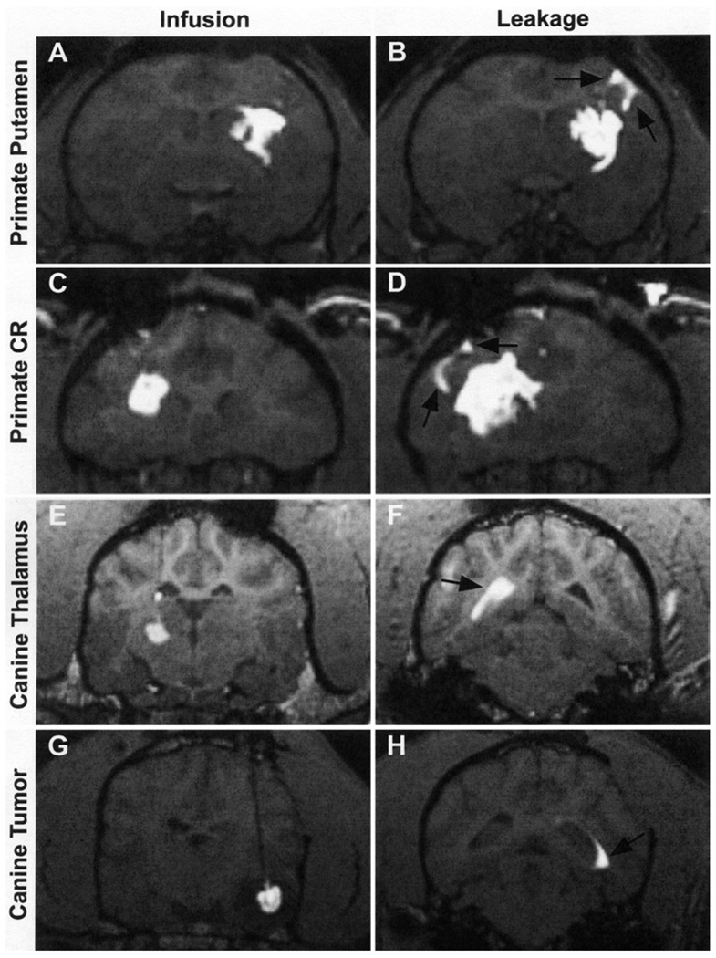
FIG. 1.
Coronal MR images showing the CED distribution of gadoteridol liposomes in 4 specific sites and the leakages associated with each infusion. A and B: Primate putamen infusion and corresponding sulcal leakage. C and D: Primate corona radiata infusion and corresponding sulcal leakage. E and F: Canine thalamus infusion and corresponding ventricular leakage. G and H: Canine tumor infusion and corresponding ventricular leakage.

