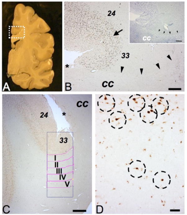
Figure 1. Gray and white matter structures surrounding cingulate cortex.
(A) Representative coronal slab of a hemisphere (postmortem), rectangle outlines approximate position and size of tissue block destined for present study. (Image courtesy of Mr. L. Fernandez, Harvard Brain and Tissue Resource Center.) (B) Parallel sections stained for NeuN immunoreactivity or Nissl (insert) showing cingulate cortex with dorsal BA24 and ventral BA33, transition marked by arrow. See text for details. The border between the cingulate white matter (including cingulum bundle) and the fiber tracts of bordering corpus callosum (cc) is demarcated by arrowheads. (C) Digitized image from region-of-interest, including the position of counting frame and white matter compartments I-V each 500 micron deep and 2000 microns wide (see Methods). * in B, C marks dorsal line of corpus callosum with induseum griseum. (D) Higher resolution image from superficial white matter bordering cingulate cortex, showing several NeuN+ neurons (marked by dotted circles), including two cells completely surrounded by WM tissue (bottom). Size bars (in μm), (B) 500, (C) 1000 and (D) 100.

