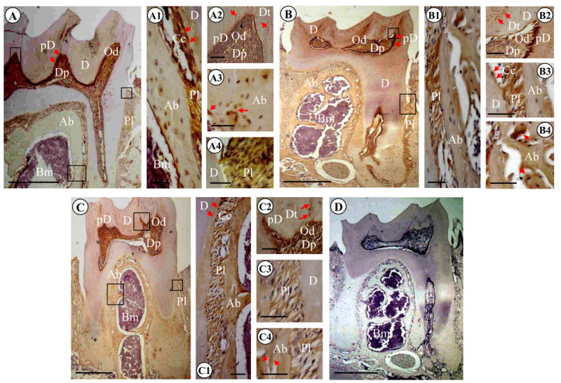
Fig. 4. Light microscopic images of mouse mandibular molars of 3.5, 7.5 and 13.5 month-old mice.

(A) 3.5 month mouse molars. Immunopositive reactions using anti-DSP antibody were observed in dentinal tubules of mantle dentin, predentin (arrows), odontoblasts and dental pulp. DSP expression was also detected in cells and ECM within alveolar bone and periodontal ligament. A1, A2, A3 and A4 are high magnifications of A. (B). 7.5 month first mandibular molar. DSP signal was seen in dentinal tubules of mantle dentin, predentin (arrows), odontoblasts and dental pulp. Positive reactions for DSP were also evident in cells and ECM within alveolar bone and periodontal ligament. B1, B2, B3 and B4 are high magnifications of B. (C). 13.5 month first mandibular molar. Immunostaining reactions for DSP were present in dentinal tubules of mantle dentin, predentin (arrows), odontoblasts and dental pulp. DSP signal was also intense in cells and ECM within alveolar bone and periodontal ligament. C1, C2, C3 and C4 are high magnifications of C. (D). 7.5 month mandibular molars. The section was stained with hematoxylin and eosin with negative control. Bar: 500 μM in A, B, C and D; 50 μM in A1, A2, A3, A4; B1, B2, B3, B4; and C1, C2, C3, C4. Ab, alveolar bone; Am, ameloblasts; Cc, cellular cementum; D, dentin; Dp, dental pulp; Dt, dentinal tubules; E, enamel; Od, odontoblasts; Pd, predentin; Pl, periodontal ligament.
