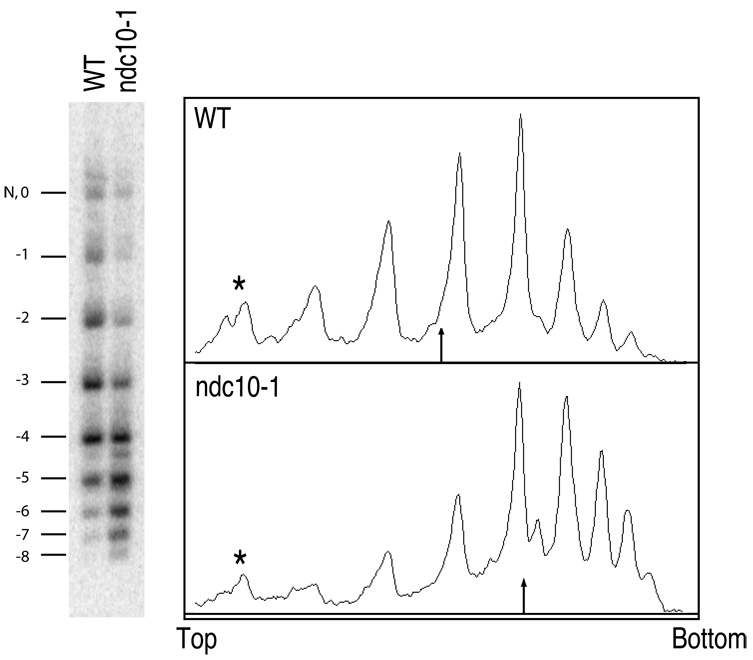
Figure 3. A yeast minichromosome loses negative supercoils with a functioning centromere.
Total DNA was isolated from either a wildtype (WT) or a mutant ndc10-1 strain carrying the same CEN3 minichromosome (Figure 2B), released from α-factor arrest at the restrictive temperature (37°C), and purified DNA was resolved on an agarose gel containing 0.3µg/ml chloroquine. A Southern blot to detect the minichromosome is shown on the left. A densitometry trace of each lane is shown on the right. The most slowly migrating band contains both a nicked circle (N) and a topoisomer with no net writhe in the presence of chloroquine (0). The numbers correspond to the value of net writhe in this chloroquine gel. The minichromosome has fewer negative supercoils in the wildtype strain with Cse4p present at the centromere, compared to the ndc10-1 mutant strain, which loses Cse4p at the restrictive temperature. The arrow in the trace indicates the location of the mean topoisomer distribution. The asterisks mark the bands containing nicked and “0” topoisomer, which was omitted from calculating the mean distribution. Nicked and ”0” topoisomer were resolved by two-dimensional electrophoresis, where similar results were obtained (Figure S4).

