
Figure 3.
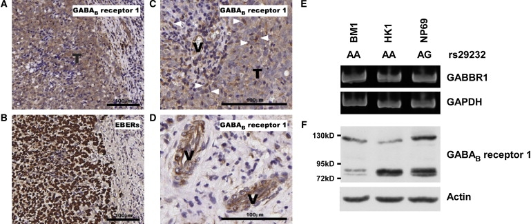
GABAB Receptor 1 Protein Expression in NPC Tumor Cells
GABAB receptor 1 protein was detected in NPC tumor cells (A), infiltrating leukocytes (C, indicated by arrowheads), and endothelial cells (D, indicated by “V”) by immunohistochemical staining. Distribution of NPC tumor cells is depicted with positive nuclear EBER signals (B). Expression of GABBR1 transcripts (E) and protein (F) was detected in NPC cell lines and in the normal nasopharyngeal epithelial cell line, NP69.