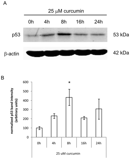
Figure 5. Curcumin induces p53 expression.
A). Cells were treated with 25 µM curcumin for the indicated time points before cells were lysed and proteins were separated by SDS-PAGE and blotted onto nitrocellulose membranes. Blots were incubated with Abs against p53 and β-actin (as a loading control) and the appropriate fluorescently labeled secondary Abs. B). Fluorescence of the specific protein bands was determined using the Odyssey Infrared Imaging System. Shown are the mean±SD of the band intensities of Hsp70 corrected for β-actin from 3 independent experiments, * = p<0.05 compared to 0 h.

