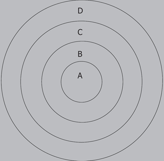
Figure 1.

A: The area of VX-2 tumor center; B: The area of VX-2 tumor periphery; C: The area of VX-2 tumor outer layer; D: The normal liver parenchyma area around tumor when the values of ADC and signals were measured on DWI and samples were investigated pathologically.
