
Figure 3.
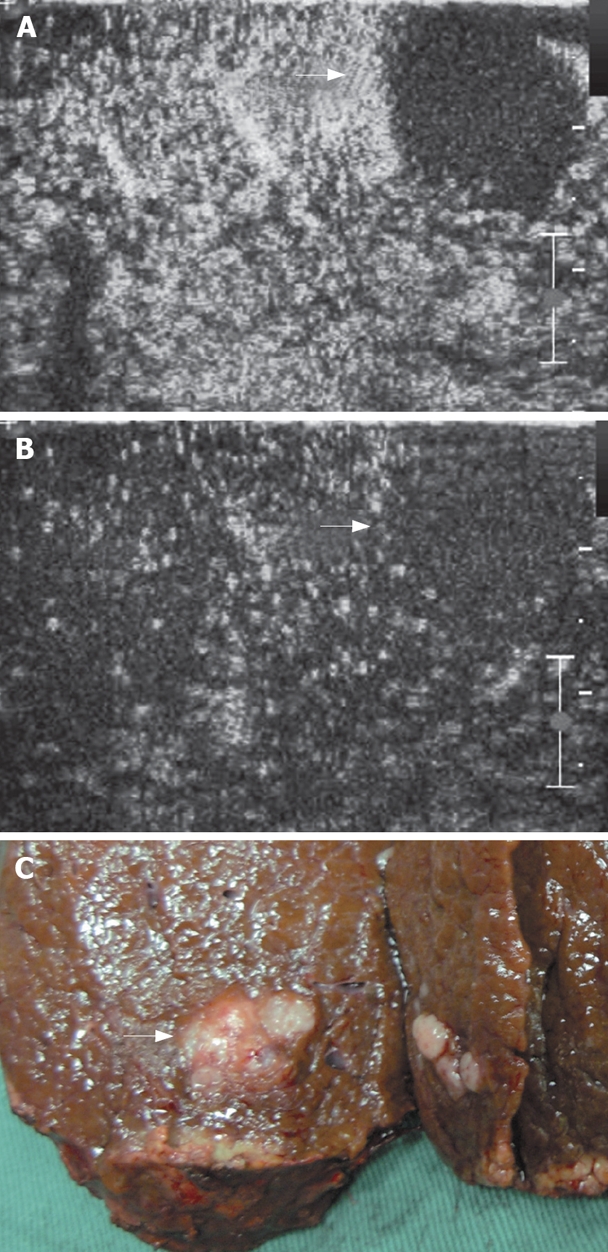
A mosaic nodule in a cirrhotic liver at IOUS. A: The nodule shows no contrast agent uptake in arterial phase (arrows); B: The nodule shows no contrast agent uptake in late parenchymal phase (arrows); C: Specimen of the mass after resection proved to be HCC at histology.