
Figure 2.
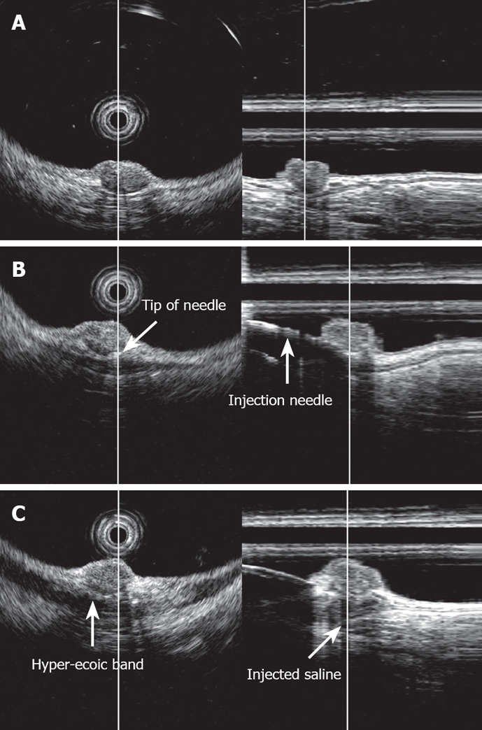
EUS image of rectal carcinoid (radial and linear image). A: Carcinoid was imaged as a low-echoic region with a depth of invasion to the third layer, i.e. the submucosal layer; B: Image upon saline injection into the submucosal layer. The tip of the injection needle was located beneath the tumor in the submucosal layer; C: Image after saline injection into the submucosal layer. The injected saline was imaged as a low-echoic area beneath the tumor in the submucosal layer.