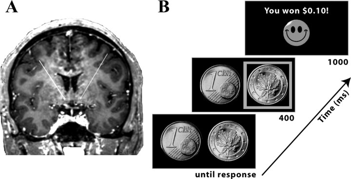
Figure 1.
A, Placement of DBS electrodes in nucleus accumbens in one patient. Displayed is an MRI from one patient used in electrode placement planning. The “X” (ventral end of the lines) shows the location of the electrode probe through this slice. B, Overview of experimental design and timing of events.

