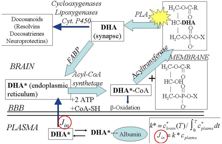
Figure 1. Model of brain docosahexaenoic acid cascade at the synapse.
Docosahexaenoic acid (DHA), esterified at the sn-2 position of a phospholipid, is liberated by activation (star) of PLA2 at the synapse, secondary to neuroreceptor activation 1, 37. A fraction of the unesterified DHA is converted to docosanoids by COX, lipoxygenase or P450 enzymes, whereas the remainder is transported by a fatty acid binding protein (FABP) to the endoplasmic reticulum. From there, DHA is activated to docosahexaenoyl-CoA by an acyl-CoA synthetase with the consumption of two ATPs, then esterified into an available lysophospholipid by an acyltransferase. Unesterified DHA also can be lost by β-oxidation in mitochondria or peroxisomes, or by other pathways (not shown). The endoplasmic reticulum compartment is in very rapid equilibrium with unesterified plasma DHA that has been dissociated from circulating albumin, whereas the synaptic compartment does not exchange with plasma DHA 70. This allows injecting radiolabeled DHA* intravenously and determining the incorporation rates Jin (circled), a critical parameter, of unesterified unlabeled plasma DHA into individual membrane phospholipids, as well as DHA turnover rates and half-lives in those phospholipids. Adapted from 35.

