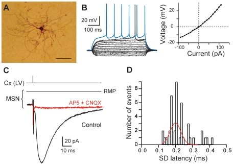
Figure 1. MSN characterization and corticostriatal monosynaptic transmission.
(A) High magnification of a MSN injected with biocytin (scale bar, 100 µm). (B) MSN membrane properties and spiking pattern: a hyperpolarized RMP, an inward rectification (illustrated in the steady-state I-V relationship) and a long depolarizing ramp to the action potential threshold leading to a delayed spike discharge. Raw traces show voltage responses to 500 ms current pulses from −90 pA to 110 pA with 20 pA steps and to +40 pA (blue trace) above action potential threshold. (C) Cortically-evoked MSN EPSCs (averages of 7 traces) in control and with CNQX (10 µM) and AP5 (50 µM). (D) Distribution of latency SD was centered on 0.20 ms and fitted by a Gaussian function. These values of SD latency indicate a monosynaptic corticostriatal transmission because inferior to 0.5 ms. Cx (LV), cortical layer V.

