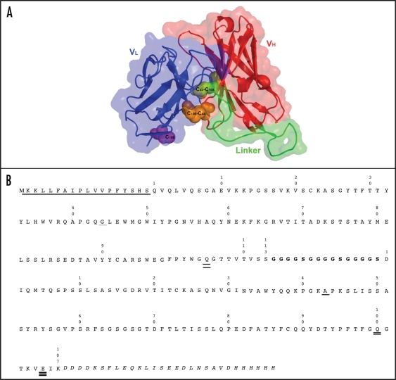
Figure 3.
Molecular model and sequence of BHA10 scFv. (A) The scFv is in the VH□VL orientation. VH is shown in red, VL is shown in blue, and the Gly/Ser linker is shown in red. Paired cysteine substitutions are shown at VH and VL positions 44 and 100, and 105 and 43, respectively. The VH 105C is shown unpaired as it utilizes VH 44C. (B) Amino acid sequence of wild-type BHA10 scFv. The Gene III signal peptide is shown underlined, (Gly4Ser)3 linker is indicated in bold type, and the Enterokinase site, Myc and His tags are indicated in italics. Positions of cysteine substitutions are as follows—the VH substitution at position 44 is shown as a single underline and at position 105 as a double underline. The VL substitution at position 43 is shown as a single underline and at positions 100 and 105 as double underlines.

