Figure 3.

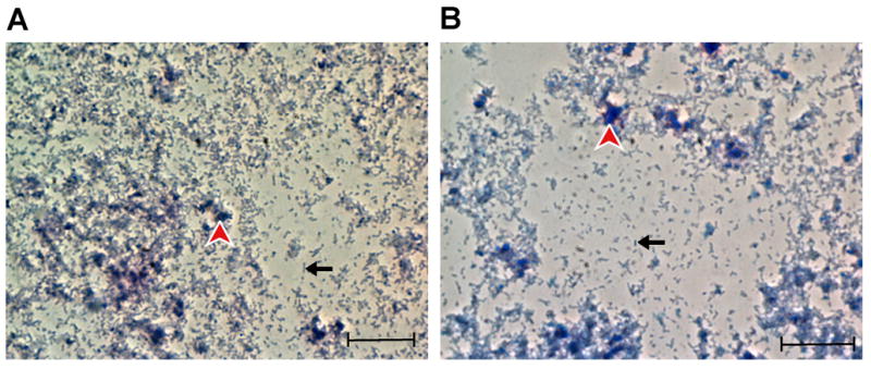
Rickettsiae, partially purified from Vero 76 cells. The black arrow points to an individual bacterium, and the red arrowhead points to cellular debris. (A) Giménez staining. (B) Diff-Quik staining. Scale bar = 10 μm

Rickettsiae, partially purified from Vero 76 cells. The black arrow points to an individual bacterium, and the red arrowhead points to cellular debris. (A) Giménez staining. (B) Diff-Quik staining. Scale bar = 10 μm