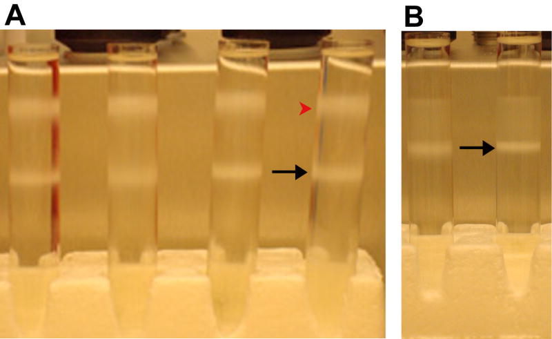
Figure 4.
Ultracentrifuge tubes with rickettsiae purified from Vero 76 cells by Renografin density gradient ultracentrifugation. The rickettsiae were purified from six 150cm2 flasks of heavily infected cells. (A) Tubes after the first run through the Renografin gradient. The black arrow points to the band of viable rickettsiae at the 32%–36% Renografin interface. The red arrowhead points to the debris at the SPG-32% Renografin interface. (b) Tubes after the second run through the Renografin gradient. The black arrow points to the band of viable rickettsiae at the 32%–36% Renografin interface.

