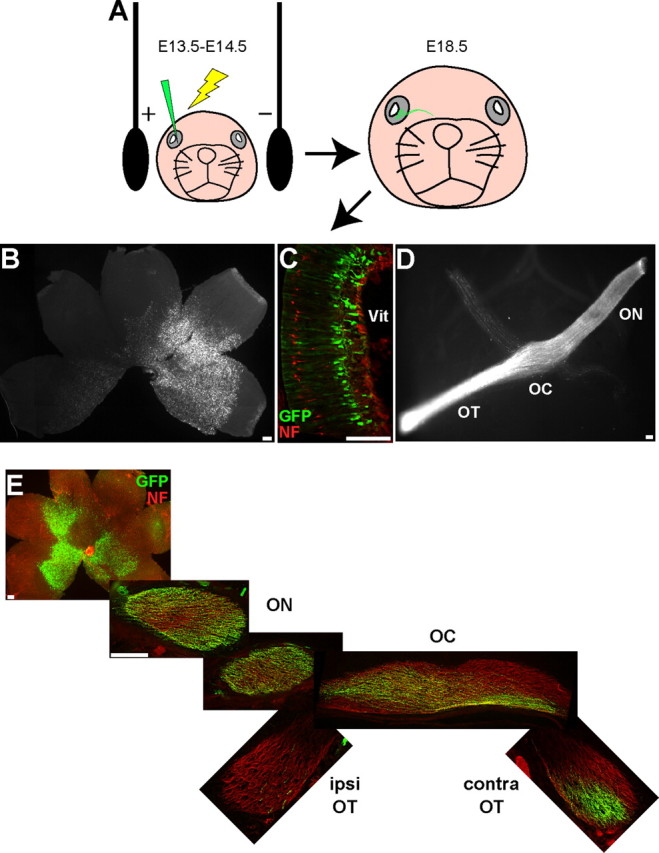
Figure 1.

GFP expression in RGCs after in utero retinal electroporation. A, Diagram depicting in utero retinal electroporation procedure. All images are taken from E18.5 embryos electroporated with GFP at E14.5. B, C, GFP+ retinal cells are clearly visible in both whole-mount (B) and cryosectioned (C) retina, where the GFP+ cells are predominantly localized to the ganglion cell layer. D, E, GFP+ axons can be visualized throughout the optic nerve (ON), optic chiasm (OC), and OT in either the semi-intact visual system preparation (D) or serial cryosections throughout the projection pathway (E). NF, Neurofilament; Vit, vitreous; ipsi, ipsilateral; contra, contralateral. Scale bars, 100 μm.
