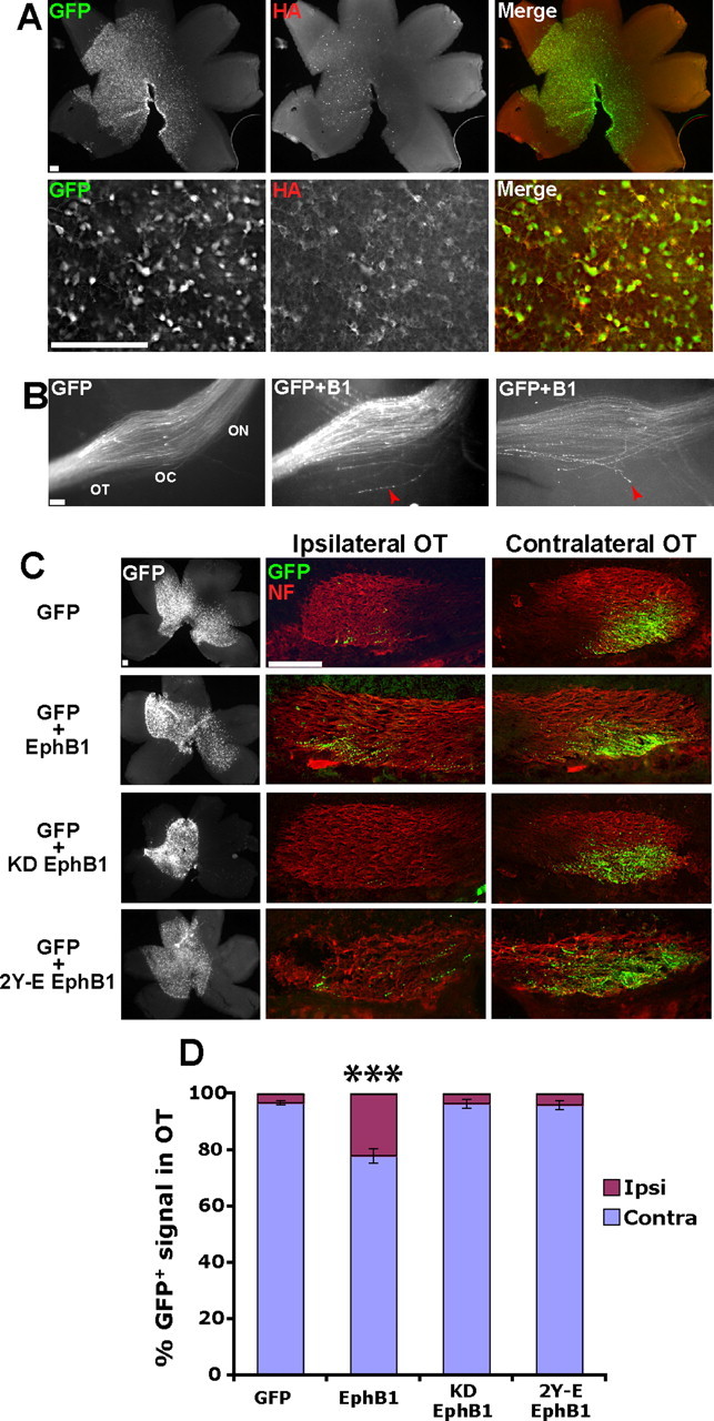
Figure 2.

Ectopic EphB1 expression converts crossed RGC projections to an ipsilateral fate in a kinase-dependent manner. A, Examples of E18.5 retina electroporated with GFP plus HA–EphB1 at E14.5. Top, Low-power view of entire retina. Bottom, Higher-power images demonstrating that the vast majority of GFP+ cells are also HA+, indicating that GFP faithfully recapitulates ectopic EphB1 expression. B, The optic chiasm (OC) in semi-intact visual system preparations of embryos electroporated with GFP (left) or GFP plus EphB1 (middle and right). Note that some GFP+ axons from GFP plus EphB1 electroporated embryos misproject posteriorly at the optic chiasm (red arrowheads), which was rarely observed in embryos electroporated with GFP alone. n ≥ 7 for each condition. ON, Optic nerve. C, Whole-mount retina and cryosections through the ipsilateral and contralateral OTs from embryos electroporated with GFP, EphB1, KD EphB1, and 2Y-E EphB1. D, A significant increase in GFP+ ipsilateral (Ipsi) projections is observed in embryos electroporated with GFP plus EphB1 compared with GFP alone (22 vs 3%). This increase is not observed in KD EphB1 or 2Y-E EphB1 electroporated embryos. n ≥ 9 embryos for each condition, from three or more separate electroporation experiments. Contra, Contralateral. Data represent mean ± SEM. Scale bars, 100 μm. ANOVA: F(3,48) = 27.09, p < 0.0001; modified t tests: ***p < 0.0001.
