Figure 7.

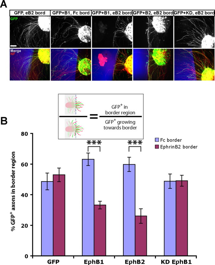
EphB2 electroporated retinal axons are repelled by ephrin-B2 substrates. A, Examples of explants from ex vivo electroporated retina plated adjacent to ephrin-B2 (eB2) or control Fc substrate borders (blue regions; see Fig. 6). NF, Neurofilament. Scale bar, 100 μm. B, GFP+ RGC axons from both EphB1 and EphB2 electroporated retina are repelled by ephrin-B2 borders, whereas RGC axons electroporated with GFP alone or KD EphB1 project uninhibited into the ephrin-B2 region. The diagram depicts method of analysis, with GFP axons in the gray region not included in calculations. n ≥ 12 explants for each condition, from at least two separate experiments. Data represent mean ± SEM. ANOVA: F(7,127) = 8.08, p < 0.0001; modified t tests: ***p < 0.0001.
