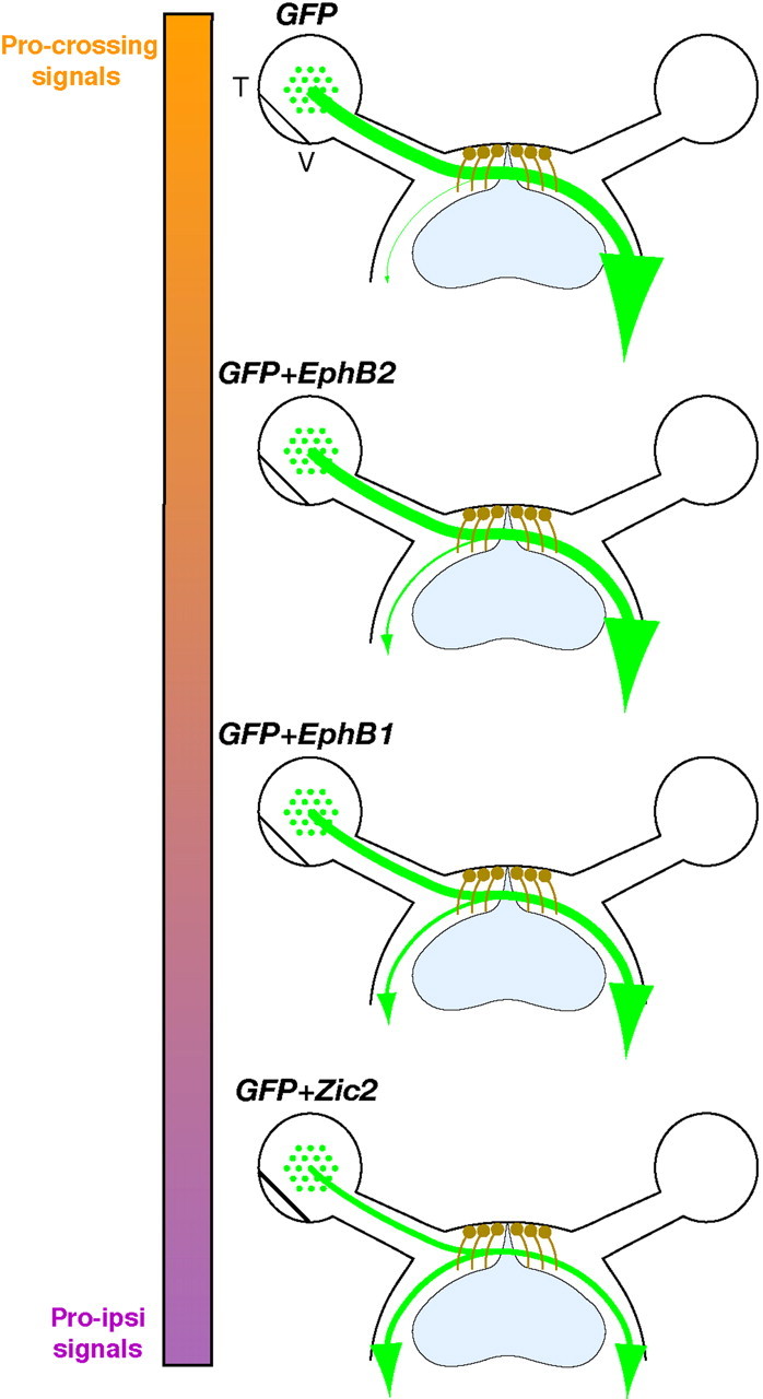
Figure 9.

Effects of ectopic gene expression on retinal axon divergence at the optic chiasm. Diagrams summarize retinal fiber projection at the optic chiasm after ectopic expression of EphB1, EphB2, and Zic2 in non-VT RGCs. Nearly all GFP+ axons cross the midline when GFP alone is electroporated into embryonic retina. A moderate increase in GFP+ ipsilateral projections is observed in retina electroporated with GFP plus EphB2 (∼10%), whereas RGCs electroporated with GFP plus EphB1 display an even greater increase in uncrossed GFP+ fibers (∼22%). The strongest effect is observed with Zic2, which is capable of converting ∼50% of non-VT GFP+ RGC axons to an ipsilateral fate. The colored bar represents theoretical relative percentages of pro-crossing (orange) and “pro-ipsilateral” (Pro-ipsi; purple) signals in GFP+ RGCs. T, Temporal; V, ventral.
