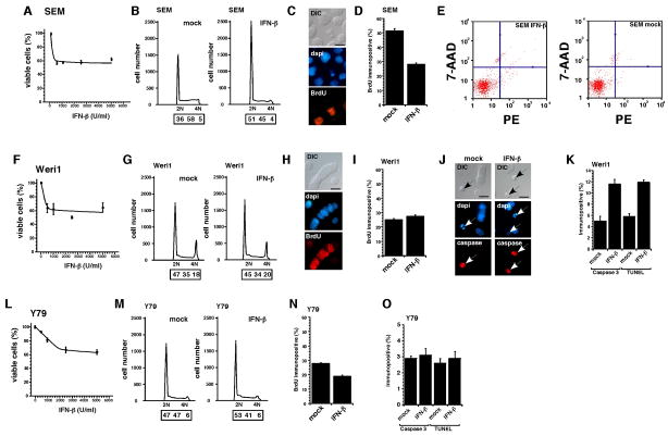
Fig 1. Retinoblastoma cell lines are sensitive to interferon-β in culture.
SEM (leukemia positive control) showed a maximum reduction in viability at 500 IU/mL of IFN-β (A). Cell cycle arrest at G0/G1 (B) and suppression of proliferation indicated by BrdU labeling (C,D) are the primary mechanisms for this response. There was no significant increase in apoptosis of SEM cells following exposure to 500 IU/ml of IFN-β. The Weri1 retinoblastoma cells showed a similar sensitivity to IFN-β with a maximum reduction in viability at 500 IU/mL (F). However, in contrast to SEM cells, the Weri1 cells exhibited no detectable change in proliferation (G–I) and a significant increase in apoptosis following exposure to IFN-β as measured by expression of activated Caspase-3 and the presence of TUNEL+ cells (J,K). Y79 retinoblastoma cells were the least sensitive cells tested in our experiments (L). Following exposure to IFN-β Y79 cells exhibited a small increase in the proportion of cells in the G1 phase (M) of the cell cycle consistent with a decrease in the proportion of BrdU labeled cells following IFN-β treatment (N). There was no significant increase in apoptosis in Y79 cells exposed to IFN-β (O). Scale bars: 10μm.

