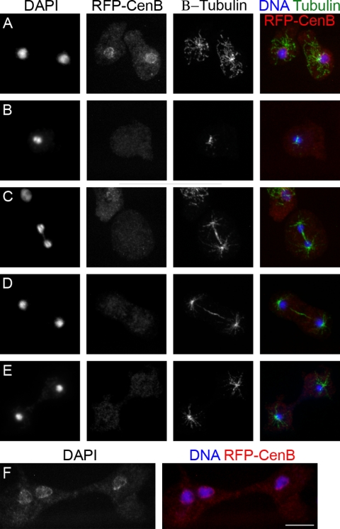
FIG. 4.
The nuclear localization of RFP-DdCenB is bound to the cell cycle. Confocal microscopy images show the loss of nuclear localization of RFP-DdCenB as cells enter mitosis and continue through to cytokinesis. A, interphase; B, early mitosis; C, late mitosis; D, early cytokinesis; E, late cytokinesis. RFP-DdCenB remains at the nuclei of cells undergoing traction-mediated cytofission (F). Images are collapsed frames from stacks of 11 slices of 0.2 μm each. Cells expressing RFP-DdCenB were fixed with chilled methanol, labeled with antibodies against β-tubulin (as indicated), and stained with DAPI. Secondary antibody: anti-mouse Alexa 488. Bar, 5 μm.

