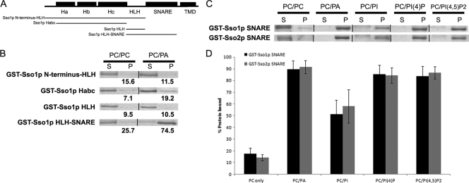
FIG. 2.
The SNARE (H3) domains of both Sso proteins mediate binding to PA and other phosphoinositides in vitro. (A) Schematic of the structural organization of Sso1, with regions fused to GST used in liposome binding assays indicated below. (B) Images of binding assays with GST fusions on SDS-PAGE gels stained with Coomassie. Liposomes were prepared in a 1:1 molar ratio with 34:1 PC and the lipid species listed above corresponding assays. Supernatant (S) and pellet (P) represent the components of a single assay. Values below images of pellet fractions are percent protein bound as determined by densitometry from individual assays (see Materials and Methods). (C) Representative images of binding assays with GST-Sso SNARE proteins; liposomes were prepared as described for panel B. (D) Graph of percent protein bound for all liposome species tested with GST-Ssop SNARE fusions. All assays were performed in triplicate; error bars represent SEM.

