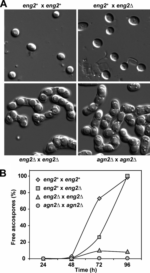
FIG. 3.
Eng2 participates in ascus wall hydrolysis following sporulation. (A) Microscopic appearance of sporulated cultures obtained from crosses between wild-type haploid (OL176/OL177), eng2+/eng2Δ (OL176/OL773), eng2Δ/eng2Δ (OL759/OL773), and agn2Δ/agn2Δ (OL763/OL777) strains. Cultures were incubated for 96 h before the images were taken. (B) Quantification of ascospore release from the asci indicated in sporulated cultures from the same crosses. At the indicated time intervals, the percentages of free ascospores were determined by light microscopy. At least 150 cells were counted for each time point.

