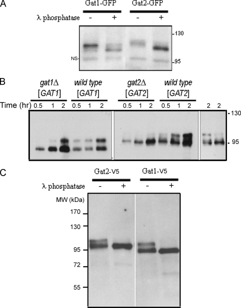
FIG. 5.
Gat1p and Gat2p are phosphoproteins. (A) Wild-type cells expressing endogenous levels of Gat1-GFP or Gat2-GFP were grown to mid-exponential phase in liquid culture and then lysed in the presence of phosphatase inhibitors, followed by subcellular fractionation, as indicated in Materials and Methods. Fifteen micrograms of protein from microsomal fraction preparations were acetone precipitated, resuspended in phosphatase assay buffer, and either left untreated or incubated with λ phosphatase (400 units) for 20 min at 30°C. Samples were then subjected to Western blot analysis with anti-GFP antibodies (as described in Materials and Methods). NS, nonspecific band. (B) Strains CMY203 (gat1Δ) and CMY201 (gat2Δ) were transformed with pGAL1-GAT1-V5 or pGAL1-GAT2-V5, respectively. Isogenic wild-type cells were also transformed with the same plasmids. Transformants were grown to mid-exponential phase in selective media containing 2% raffinose (time zero), and then expression was induced by shifting the cells to fresh media containing 2% galactose. Samples were collected at the indicated time points after the shift and lysis was performed in the presence of phosphatase inhibitors. Expression of Gat1p and Gat2p (5 μg of total protein) was analyzed by Western blotting using anti-V5 antibodies. (Right) Gat2p pattern in wild-type and gat2Δ cells after 2 h of the shift. Deliberately less total protein was loaded for the wild-type sample to show that the differences observed in the pattern are not due to differences in total Gat2p present in the sample. (C) Cell extracts from gat1Δ(pGAL1-GAT1-V5) and gat2Δ(pGAL1-GAT2-V5) transformants similar to the ones used in panel B were grown on galactose to mid-exponential phase and subjected to immunoaffinity precipitation using anti-V5-agarose beads. Immunoprecipitates were washed, as indicated in Materials and Methods, and either left untreated or incubated with λ phosphatase (200 units) for 30 min at 30°C. Beads were then subjected to Western blot analysis with anti-V5 antibodies. MW, molecular weight.

