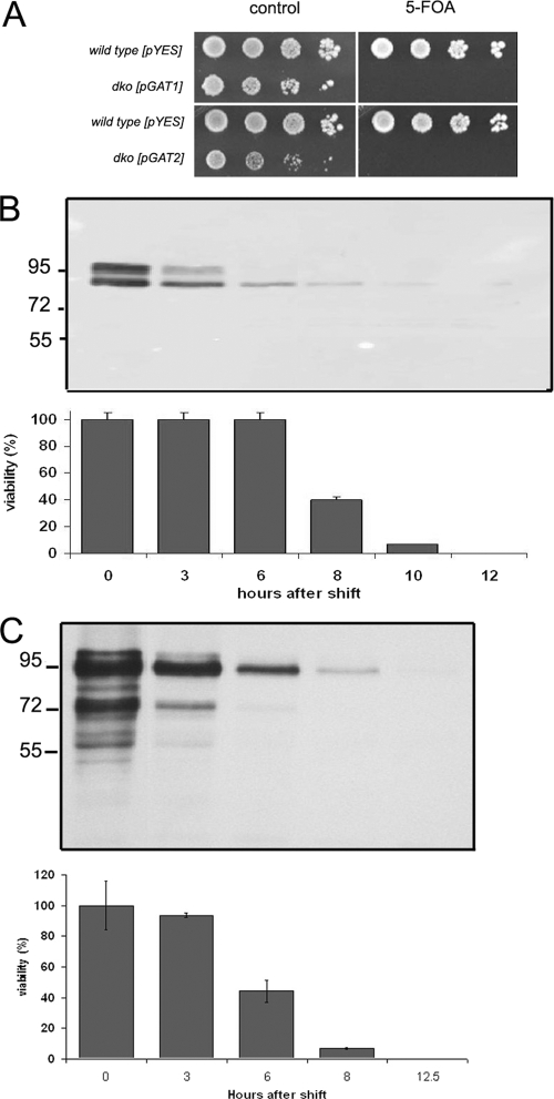
FIG. 6.
Characterization of gat1Δ gat2Δ double-knockout strains. (A) Strains simultaneously lacking GAT1 and GAT2 but maintained by using (pGAL1-GAT1-V5, URA3) dko-GAT1 or (pGAL1-GAT2-V5, URA3) dko-GAT2 were grown on media containing 2% galactose. Cultures were serial diluted and plated on galactose-defined medium lacking uracil (control) or same medium containing 5-fluoroorotic acid (5-FOA). (B, C) Isogenic wild-type strains transformed with the empty pYES-URA3 vector were used as a control. dko-GAT1 (B) or dko-GAT2 (C) cells were shifted to glucose-containing medium, and samples were collected at the indicated time points after the shift. (Top) Western blot analysis of 10 μg total protein analyzed for each time point. (Bottom) Percentage of viable cells during the time course, performed as indicated in Materials and Methods. A 55% and 38% reduction in the levels of Gat1p and Gat2p, respectively, was estimated 3 h after the shift (while cells are fully viable).

